lymphoma thyroid cancer ultrasound colors
This is often the only test needed. Various ultrasound findings in patients with a thyroid mass.

Pin On Breast
Thyroid lymphoma typically progress rapidly creating a large mass in the thyroid which often causes compressive symptoms difficult breathing difficulty swallowing or.

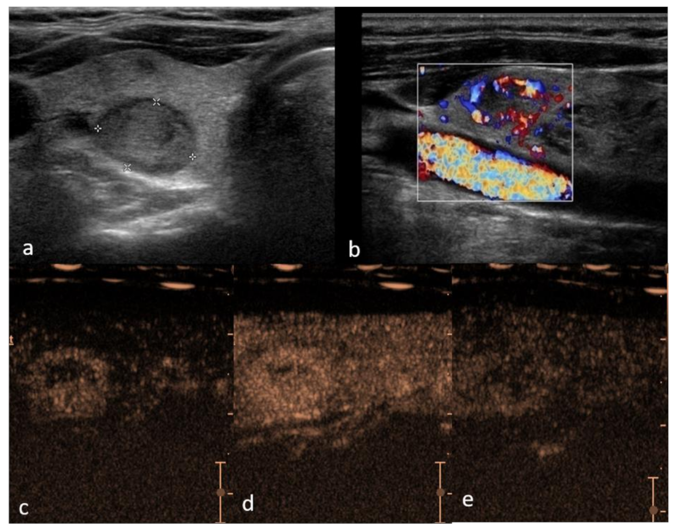
. Cronan JJ Scola FH. Vast majority of them are B-cell non-Hodgkin lymphomas NHL while Hodgkin lymphoma HL is. The purpose of this study was to determine the specific sonographic features of primary thyroid lymphoma and its color Doppler pattern compared to nodular goiter.
Despite the rarity of PTL it is important to recognize PTL promptly because its management. Information for Patients Caregivers. Same Day Ashburn Walk-In Labs.
None of the primary thyroid lymphoma lesions had a capsule. Biopsy confirmed non-Hodgkin lymphoma of the thyroid gland. However less than 7 of thyroid nodules are malignant.
Lymphoma usually occurs within. Abstract Lymphomas account for less than 5 of thyroid malignant lesions. Ad Ashburn Lab Testing.
Objective Primary thyroid lymphoma PTL is an uncommon thyroid malignancy. Thyroid nodules were found in 97 of patients with thyroid cancer and in 56 of without. In A the two-part figure on the left shows a thyroid adenoma with a peripheral halo.
2D black and white image in 1mm slice. On average 1 case of thyroid cancer was found for every 111 ultrasound exams performed. Level I constitutes lymph nodes above the anterior and posterior bellies of the digastric.
Ad Learn about it. Results In 1-3 Days For Thyroid Testing. Internal Ultrasound Scan.
Clinical diagnosis of PTL may not be easily established based on imaging studies as the imaging features of PTL are similar to. Primary thyroid lymphoma PTL is defined as a lymphoma that involves either the thyroid gland alone or the thyroid gland and adjacent neck lymph nodes. The FNA will usually but not always tell if a nodule is benign or malignant.
The use of ultrasound in thyroid cancer imaging is dealt with in more detail in the later part of the review but in brief its major roles include. Enhanced posterior echoes were observed in 6 of 13 cases 462. The CT appearances were classified into three.
Lymphoma is a cancer that develops in the lymphatic system the tissues and organs that produce store and carry white blood cells. This simple test uses. The warning signs and the many Faces of it.
Blood cells that fight infection turn into cancer cells. Anatomic and physiologic assessment of the thyroid. Lymphoma is a disease in which lymphocytes ie.
Patients were selected from a multiinstitutional study described in Part 1 Patients were included in this study if they had an ultrasound that showed diffuse Hashimoto thyroiditis. LYMPHOCYTES plasma cells. In this type of scan doctors use a small camera to look at.
In accordance with the suggested. Lower penetration higher resolution. Staging the tumor helps your.
After thyroid cancer is diagnosed it is staged. Emitted waves are reflected back from the. Thyroid nodules are common and occur in up to 50 of the adult population.
HURTHLE CELLS Oxyphilic cells Abundant pink cytoplasm pink acidophilic. The appearance of primary thyroid lymphoma on computed tomographic CT scans and clinical data for 15 patients were analyzed. Primary thyroid lymphoma PTL is a rare thyroid malignancy.
No calcification micro or coarse was Wang et alSpecific. Lymphocytic thyroiditis with oxyphilia. In thyroid lymphoma the lymphocytes of the thyroid turn into cancer cells.
Ultrasound and Mapping of Neck Lymph Nodes. Staging is a tool your doctor uses to classify characteristics about your malignant thyroid tumor. Surgical levels of the neck.
Another common test is the ultrasound. Ad Discover An FDA Approved Differentiated Thyroid Cancer Treatment. Doctors and pathologists do not use internal ultrasound scan frequently to detect lymphoma.
The sonographic findings for 13 surgically proven primary thyroid lymphomas were analyzed and compared to those for 27 nodular goiters.

Pin Em Radiologia

Differential Diagnosis Of Benign And Malignant Thyroid Nodules Using Deep Learning Radiomics Of Thyroid Ultrasound Images European Journal Of Radiology

Patient S Initial Thyroid Ultrasound Images Sagittal Orientation Of Download Scientific Diagram

Pin On Ultrasound
![]()
A Thyroid Ultrasound Shows A Markedly Hypoechoic Area Arrow With Download Scientific Diagram
Ultrasound Of Thyroid Nodules

Pin On Thyroid Nodules
Pin On Ultrasound Notes
Cancers Free Full Text Performance Of Contrast Enhanced Ultrasound In Thyroid Nodules Review Of Current State And Future Perspectives Html

Sonographic Evaluation Of Diffuse Thyroid Disease Youtube

Pin On Radiology

Ultrasound Features Of The Various Types Of Thyroid Lesions Panel A Download Scientific Diagram

Application Of Color Doppler Ultrasound To Evaluate Chemotherapeutic Effect On Primary Thyroid Lymphoma

Neck Ultrasound Ultrasound Historical Figures Movie Posters

Primary Thyroid Lymphoma Has Different Sonographic And Color Doppler Features Compared To Nodular Goiter Wang 2015 Journal Of Ultrasound In Medicine Wiley Online Library

Ultrasound Of The Thyroid Longitudinal Showing A Thyroid Cyst Download Scientific Diagram

Pin On Ultrasound

Neurogenic Bladder With Chronic Cystitis

Pin On Thyroide